advantage:
Fully automated image acquisition and analysis
Quickly get multiple phenotypic parameters for each cell
Real-time monitoring of the toxicity of living cells, which can last from a few minutes to a few days
The nervous system is very sensitive to many toxic compounds and harmful substances produced in the natural environment. During the course of the disease, neurotoxicity can cause transient or persistent damage to the brain or the peripheral nervous system, such as spinal cord injury, stroke, traumatic brain injury, and the like. At the same time, this neurotoxicity is also the main cause of many neurodegenerative diseases such as Alzheimer and Parkinson.
High-throughput imaging techniques that induce human pluripotent stem cells can be used to screen for neurotrophic, protective or neurotoxicity of drug candidates or environmental pollutants. This section explains how to combine iPSCs, molecular instruments and software to automatically screen endpoints for pluripotent stem cells and neurotoxicity of live cells.
iPSC-based toxicity screening
Induction of pluripotent stem cell technology is very useful for our study of neuronal toxicity, and iPSCs can display the function of mature neurons in large quantities. By combining this unique biologically relevant cell type with high-throughput imaging analysis, we can quickly screen for compounds that play a leading role in neurotoxicity and greatly reduce preclinical studies and related animal experiments. the cost of. What is used in this article is the fully differentiated human nerve cells of Cellular Dynamics International.
Quantify intact neural cell network
High-throughput analysis is a quantitative analytical method that measures the synaptic length to characterize the positive or negative effects of a test substance. iPSC-derived neuronal cells can form a neural network in 96 or 384 well plates within 3-5 days and then stimulate with toxic compounds for 48-72 hours. Once the neural network is labeled with ß-tubulin, images can be acquired with the ImageXpress® Micro spacious imaging system. And anti-antibody we can visualize the neural network. Relevant images and experimental data were analyzed using the Neurol Outgrowth module and AcuityXpressTM high content data analysis software in MetaXpress® image processing software.
Whether or not nuclear counterstaining is performed, the Neurolite Outgrowth module can find neuronal cell bodies and label them as positive cells based on these fluorescently labeled neural cell axons. The neural network has several important characteristic parameters including the number of axons, the length of the axons, and the number of branches per cell or region. We can calculate and count the relevant data and cell phenotype for each well.
Upper left: The results of the Neurite Outgrowth module for control and high-dose drugs analyzed the results, blue for the nucleus and green for the ß-tubulin III-labeled synapse. Upper right: Total synaptic response curve of toxic compounds. Bottom: IC50 value calculated from the total synaptic measurement response curve.
Detection of mitochondrial membrane potential
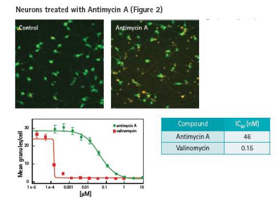
Top: Control and antimycin A treated neuronal cell images, cells were stained with JC-10. The image shows a 20x mirror shot. Bottom: The measurement response curve is plotted based on the average mitochondria/particle ratio ratio per cell.
The depolarization process of mitochondrial membrane is considered to be an important early marker of hypoxia injury and cytotoxicity. The mitochondrial membrane potential can be monitored by JC-10 dye. In normal cells, JC-10 accumulated in mitochondria is orange-red, and when the mitochondrial inner membrane potential is lowered, JC-10 is released from the mitochondria into the cytoplasm in a monomeric form and emits green fluorescence. The nerve cells were stained with JC-10 and treated with antimycin or aminomycin for 30 min. The latter two can effectively block the oxidative respiration process and calcium ion overload of the cells. Users can customize the Granularity module in MetaXpress Software to identify the density and size of JC-10 in mitochondria. Data results will be normalized to each field of view.
Detection of viable cell toxicity
Using Calcein AM and Hoechst nuclear dyes we can test the toxicity of living cells. The ImageXpress Micro system provides excellent control of the temperature, CO2 concentration, and humidity of the culture plate to accurately detect compounds that are neurotoxic (Figure 3). By comparing the neurotoxic compounds detected in the body with clinical data, we can predict the neurotoxicological properties of the compounds.
Calcein AM can simultaneously stain live nerve cell bodies and external nerve fibers. The Neuroc Outgrowth application module is used to analyze neural network networks, and the Cell Scoring application module distinguishes between living and dead nerve cells.
Top: The picture shows the toxic effects of different concentrations of methyluric on neuronal cells. The presence of Calcein AM indicates live cell metabolic activity (green). The left: IC 50 curve toxicity of the compound, based on the drawn synaptic toxicity and nuclear size measurement display. Bottom right: Hoechst-stained nuclei are marked in red.
The perfect solution for neurotoxicity screening
Molecular Devices' ImageXpress Micro system, MetaXpress software and AcuityXpress software perfectly solve the problem of how to screen for the evaluation of neurotoxic compounds. The combination of live cell screening technology and sophisticated phenotypic imaging analysis technology can effectively identify neurotoxic compounds, providing strong support for early drug development.
Natural plant intermediates,Organic Flax Seed,Organic Apricot Kernel, Organic Pine Nut,Acacetin Supplement,Piperine Blood Pressure


